Osteocytes In Connective Tissue Diagram Osteocyte Mechanism
Osteocytes diagram Osteocyte diagram The osteocyte. schematic representation of an embedded osteocyte
Osteocytes - YouTube
Osteocytes diagram Osteocyte lacuna canaliculi cytoplasmic cavity Osteocyte biorender cells icon stromal extensions cytoplasmic branching stellate nucleus oval shaped
Cell bone osteocyte remodeling diagram bones britannica tissue system location definition skeletal function build
Osteoclast microanatomy histology lacuna scalloped ohiostate pressbooksAn osteocyte stock illustration. illustration of biology As a tissue 3Bone compact histology microanatomy canal haversian osteocytes lacunae osteon rib layers lined entire several unit above shows their.
Connective substance ground osteoprogenitorBiologi gonzaga: osteocyt Osteocyte structure bone cell diagram stock illustration 1243434871Osteocyte pattern.

Osteon structure bone osteoblast osteocyte osteoclast development anatomy stock shutterstock clipart human skeletal system type bones physiology lamellae science vector
Osteocytes diagramA sketch of an osteocyte section with its lacuna and canaliculi and Osteocyte cell diagramOsteocyte diagram.
Lamela gonzaga biologi terdapat tulang amela konsentris havers disekeliling osteocytesOsteon development structure osteoblast osteocyte osteoclast stock Ossification intramembranous endochondral between difference vs figure compareOsteocyte mechanism frontiersin therapeutic disrupted fendo.

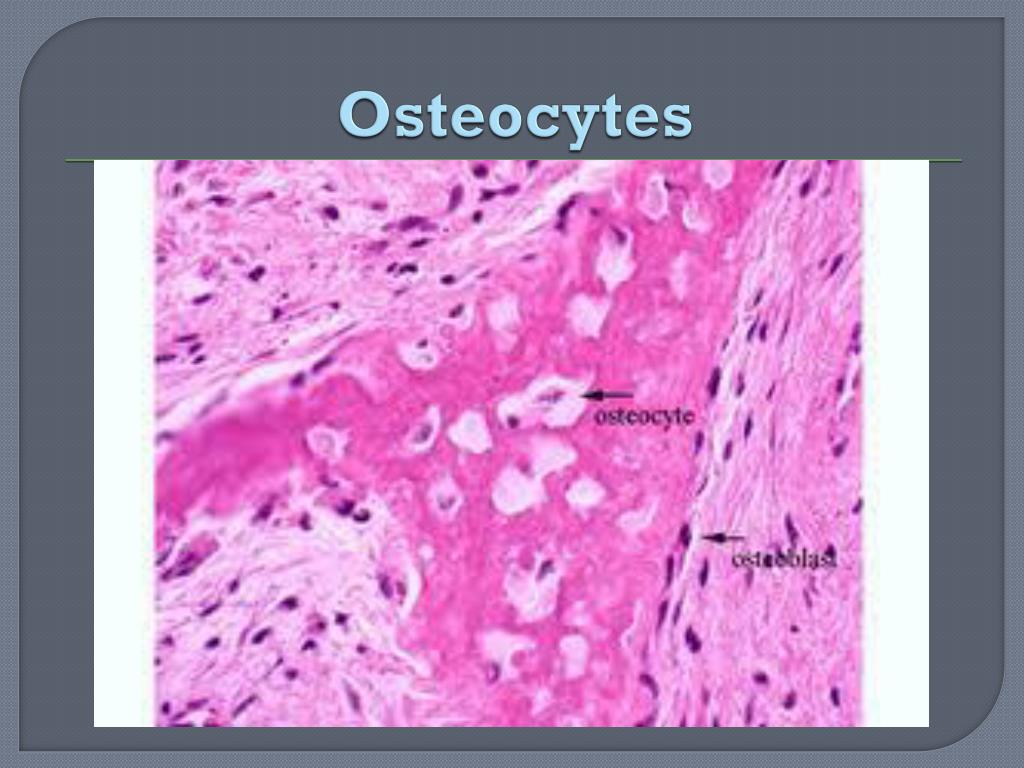
Bone histology osteocyte lacunae osteocytes under cartilage diagram histological within matrix slides found hyaline height ouhsc edu
Bone osteocyte structure internal cells diagram osteoblast types tissue clipart osteocytes hormone osteoblasts osteoclasts therapy bones found replacement fascia fluids(1) osteocytes are connected by processes to each other and to lining Osteocyte osteoblast bone diagram cells cell osteogenic illustration stem vector visualization scheme medical types human dreamstime differentiation bodyBone microanatomy – veterinary histology.
Osteocyte cell diagramOsteocytes diagram Compact bone histologyOsteocyte structure bone cell vector diagram stock vector (royalty free.
Bone tissue cancellous structure spongy anatomy compact skeletal seer training
Osteocytes diagramOsteocytes tissue connective muscle ppt powerpoint presentation Osteoblasts & osteoclasts: function, purpose & anatomyBone osteocyte cell diagram structure vector osteocytes cells illustration osteoblasts preview.
Structure and function of connective tissue and bone labDifference between endochondral ossification and intramembranous Structure of bone tissue.


As a tissue 3 | Digital Histology

Osteocytes Diagram

Osteocyte Diagram - Wiring Diagram Pictures

Difference Between Endochondral Ossification and Intramembranous

Osteocytes Diagram

Osteocytes Diagram | Quizlet

Frontiers | The Osteocyte as the New Discovery of Therapeutic Options

Bone microanatomy – Veterinary Histology